explains team lead Dr. Claire Chalopin. “Thanks to the technical expertise of ICCAS and its industry connections, practical applications based on our work are making a real-world impact in operating rooms.”
Case Study
Case Study
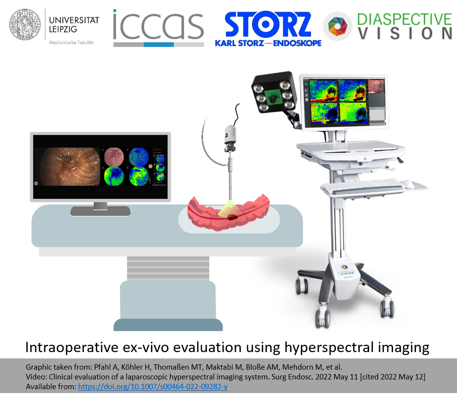
During minimally invasive surgery, cutting-edge imaging technology can give physicians more and better visual information to base decisions on. Medical spectral imaging makes the invisible visible to surgeons – and can improve outcomes for patients.
This R&D team is taking medical spectral imaging to the next level: Their computer-aided surgery solutions are non-invasive and easy to use. A combination of live video and hyperspectral images (HSI) allows surgeons to assess tissue perfusion and identify risk structures in real time during an operation.
Leipzig is home to a wide variety of highly educated medical and technology experts. They laid the groundwork for the spectral imaging success story, together with these industry-defining players:
In addition, collaboration with the two German corporate partners was made easy thanks to Leipzig’s central location and excellent infrastructure – proving that this vibrant metropolis has much to offer young ventures.
explains team lead Dr. Claire Chalopin. “Thanks to the technical expertise of ICCAS and its industry connections, practical applications based on our work are making a real-world impact in operating rooms.”
The team has helped develop technologies such as the TIVITA® Mini, the world’s first certified endoscopic spectral imaging device. By combining color video and spectral imaging, it can help improve surgical decision-making and thus enhance patient outcomes.
(Hyper)Spectral Imaging
The Leipzig University-based team of researchers and physicians has been working to advance medical spectral imaging since 2019. They collaborated with ICCAS and corporate partner Diaspective Vision GmbH in the development of the endoscopic spectral imaging device TIVITA® Mini, which is marketed by KARL STORZ SE & Co. KG. A software platform for the visualization and analysis of the spectral images is pending. More Info: www.iccas.de/projekte/hsi/
robotics
Robotic hand with various joints and cables which holds a scalpel
robotics
Robotic hand with various joints and cables which holds a scalpel
Autonomously driving minibusses, real-time transmission of ultrasound data from the ambulance…
robotics
Robotic hand with various joints and cables which holds a scalpel
With its locations in Dresden and Leipzig, the ScaDS.AI Dresden/Leipzig combines the excellent AI…
robotics
Robotic hand with various joints and cables which holds a scalpel
The great potential of artificial intelligence (AI) and machine learning for the life sciences…
robotics
Robotic hand with various joints and cables which holds a scalpel
Every human body is different, which means there is no such thing as a ‘standard’ procedure during an operation. Next3D GmbH is advancing the field of individualized surgery through augmented reality (AR) and patient-specific 3D-printed implants and instruments.